
Exploring the Potential of EEG Biomarkers in Early Alzheimer’s Disease
- 1 day ago
- 8 min read

Early detection and characterisation of Alzheimer’s disease (AD) remain major challenges in neuroscience and clinical research. As disease-modifying therapies become available, identifying subtle changes as early as possible—and understanding which individuals are most likely to progress—has become increasingly important.
At Starlab, we are interested in how advanced EEG methodologies can help us better understand the functional changes taking place in the brain during the earliest stages of cognitive decline.
In our recent preprint, “EEG Biomarkers for ATN Classification in Early Alzheimer’s Disease”, we investigated whether a structured EEG assessment could identify neurophysiological signatures associated with different stages of early cognitive decline, and how these signatures relate to established Alzheimer’s disease biomarkers.
Working together with colleagues at ACE Alzheimer Center Barcelona, we studied EEG activity in individuals with subjective cognitive decline (SCD), mild cognitive impairment without evidence of amyloid and tau pathology (MCI−), and mild cognitive impairment with an Alzheimer’s-related biomarker profile (MCI+).
Our results suggest that EEG can capture meaningful changes across these stages of cognitive decline. Importantly, they also highlight that EEG and fluid biomarkers may provide different—and potentially complementary—information about the early stages of Alzheimer’s disease.
Looking at the brain from different angles
One of the main objectives of our study was to move beyond the traditional use of resting-state EEG alone.
Alzheimer’s disease affects multiple aspects of brain function, including attention, working memory, neural synchronisation, cortical excitability and large-scale communication between brain regions. We therefore wanted to explore whether combining different EEG paradigms could provide a more comprehensive picture of these changes.
For this purpose, we used our COGBAT (COGnitive BATtery), a structured EEG battery combining four complementary paradigms:
N-back, to investigate working memory and cognitive load;
Auditory oddball, to assess attention and the brain's response to novel and relevant stimuli;
40 Hz auditory steady-state response (ASSR), to probe neural synchronisation and cortical network integrity; and
Resting-state EEG, to characterise spontaneous brain activity, including oscillatory dynamics, connectivity and signal complexity.
This combination allowed us to extract a wide range of EEG features, including event-related potentials, spectral power, functional connectivity and measures of signal complexity.
We included 60 participants, equally distributed across three groups: 20 individuals with subjective cognitive decline (SCD), 20 individuals with mild cognitive impairment without evidence of amyloid and tau pathology (MCI−), and 20 individuals with mild cognitive impairment with positive amyloid and tau biomarkers (MCI+).
The MCI groups were classified using cerebrospinal fluid biomarkers, providing an ATN-informed framework for investigating how EEG signatures relate to the underlying biological processes associated with Alzheimer’s disease.
What did we find?
The results across the four paradigms revealed a complex picture.
Rather than observing a simple, progressive deterioration of EEG measures from SCD to MCI− to MCI+, we found evidence that different neurophysiological systems may change at different stages of the disease process.
Some EEG features were associated with amyloid and tau pathology, while others appeared to be more closely related to the transition from subjective cognitive complaints to measurable cognitive impairment.
This was particularly evident when we looked at the results from each component of the COGBAT battery.
1. N-back: changes in working memory and cognitive processing
The N-back task is a working-memory task in which participants have to remember and identify information presented a certain number of steps earlier. [Find a detailed description of the task here.]
The N-back task allowed us to investigate how the brain responds when participants need to maintain and manipulate information in working memory.
Our results showed differences in event-related EEG responses associated with working memory processing across the groups. The N450 component, associated with working memory processes, showed lower amplitude in participants with MCI compared with the SCD participant (see Figure 1). These findings suggest that the neural mechanisms supporting cognitive processing under load are already altered during the early stages of cognitive decline.

In particular, ERP features extracted during the task provided information related to cognitive status and were associated with the presence of Alzheimer’s-related pathology.
This is important because task-based EEG gives us the opportunity to observe the brain while it is actively performing a cognitive operation, rather than only measuring spontaneous activity.
The N-back results therefore contribute to the broader picture emerging from our study: EEG can capture alterations in the neural processes underlying cognition, and these alterations may provide information that is complementary to conventional cognitive assessments.
2. Auditory oddball: altered attention and novelty processing
The auditory oddball task presents participants with a sequence of sounds and asks them to detect or respond to infrequent, unexpected target sounds among more frequent standard sounds. [Find a detailed description of the task here.]
The auditory oddball paradigm provided a complementary view of cognitive function by examining how the brain responds to relevant and unexpected auditory stimuli.
We observed a progressive, linear decrease in the amplitude of the P300 component across the clinical groups, from SCD to MCI− and finally to MCI+, with MCI+ participants showing the lowest P300 amplitude. The P300 component is associated with attentional processing and stimulus evaluation (see Figure 2).

Importantly, P300 amplitude was associated with both MCI+ status and CSF amyloid and tau biomarkers, suggesting that alterations in attentional processing may be linked to the underlying pathological changes associated with disease progression.
These findings support the potential of EEG-derived ERP measures to capture subtle changes in the brain's ability to efficiently process and respond to relevant information, even during the early stages of neurodegenerative disease.
Together with the N-back findings, the auditory oddball results suggest that task-based EEG may provide a valuable window into the functional consequences of early brain changes, helping to elucidate how underlying pathological processes translate into alterations in cognitive processing.
3. 40 Hz ASSR: evidence of altered cortical excitability and synchronisation
The 40 Hz auditory steady-state response (ASSR) task uses a rhythmic auditory stimulus presented at 40 Hz to investigate how effectively the brain synchronises its neural activity with an external rhythm. [Find a detailed description of the task here.]
This paradigm is particularly relevant to the study of Alzheimer’s disease because neural synchronisation and oscillatory activity are thought to play an important role in communication between brain regions and in the coordination of cognitive processes.
One of the most striking findings emerged in the MCI− group.
Despite having mild cognitive impairment, these participants did not show the same amyloid and tau biomarker profile as the MCI+ group. However, their EEG responses revealed a pattern consistent with increased cortical excitability or hyperexcitability (see Figure 3).
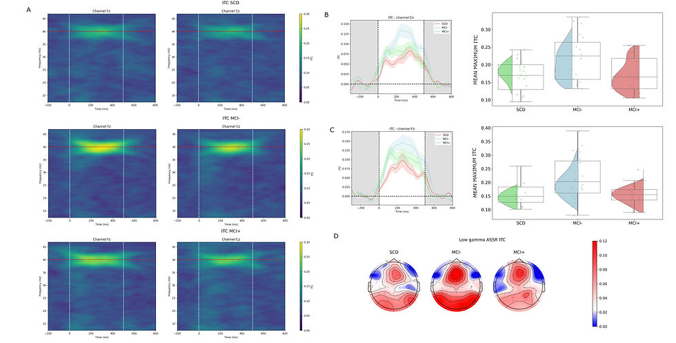
This was particularly interesting because it suggests that the relationship between cognitive impairment and neurophysiological changes may not be linear.
In other words, the brain does not necessarily move from "normal" activity to progressively "abnormal" activity in a simple, step-by-step manner.
Instead, during certain stages of cognitive decline, the brain may temporarily enter a state of increased excitability or altered network dynamics. This could potentially reflect compensatory mechanisms, an early stage of network dysfunction, or another transient neurophysiological state.
At this stage, we cannot determine exactly what the hyperexcitability pattern represents. Longitudinal studies will be particularly important to establish whether it is a transient response that precedes further changes, a compensatory mechanism, or a stable characteristic of a subgroup of individuals with MCI.
Nevertheless, this finding provides an important clue about the complexity of the neurophysiological trajectory associated with cognitive decline.
4. Resting-state EEG: oscillatory slowing, connectivity and signal complexity
In resting-state EEG, participants simply relax while their brain activity is recorded, without performing a specific task. [Find a detailed description here.]
Our resting-state EEG analyses provided another important component of the picture.
We observed differences in spectral activity, connectivity and signal complexity across the groups.
Several features were associated with amyloid and tau biomarkers, supporting the idea that spontaneous brain activity can reflect aspects of the underlying biological processes associated with Alzheimer’s disease.
In particular, measures related to oscillatory slowing provided information about disease stage and pathology. Changes in spectral power and the balance between slower and faster oscillations were among the EEG features that contributed to distinguishing the clinical groups.

We also found differences in signal complexity, providing additional evidence that the organisation and dynamical properties of brain activity change across early cognitive decline.
These findings are consistent with the broader literature showing that Alzheimer’s disease is associated with alterations in oscillatory dynamics, network connectivity and the complexity of neural signals.
However, our results also reinforce an important point: these changes do not necessarily progress in a straightforward linear fashion. The combination of resting-state and task-based measures suggests that different neurophysiological systems may be affected at different points in the disease trajectory.
Could EEG become a first step in the diagnostic pathway?
These findings lead us to an interesting question: could EEG have a role as an early, non-invasive screening tool in memory clinics?
Our results suggest that this is a possibility worth exploring.
Rather than replacing blood-based or CSF biomarkers, EEG could potentially be used as a first-pass assessment to identify individuals who may benefit from further investigation.
For example, a person presenting with subjective cognitive complaints could undergo a relatively accessible EEG assessment. If the EEG indicates patterns associated with cognitive impairment, the individual could then be prioritised for more specific biomarker testing.
This could potentially help optimise the use of more invasive or costly procedures, while providing clinicians with additional information about the functional state of the brain.
The potential value of this approach may become increasingly relevant as disease-modifying treatments continue to develop and the need for early and accurate patient stratification grows.
In future work, we need to better understand the trajectory of the hyperexcitability pattern observed in MCI− and determine whether it represents a transient compensatory response, an early stage of network dysfunction, or a stable neurophysiological phenotype.
Towards a multimodal approach to Alzheimer’s disease
For us at Starlab, this work represents an important step in exploring how EEG can contribute to the increasingly multimodal landscape of Alzheimer’s disease research.
The future of early detection is unlikely to rely on a single biomarker.
Instead, we believe that combining information about molecular pathology, cognitive performance and brain function may provide a more complete understanding of where an individual is along the trajectory of cognitive decline.
Our findings suggest that EEG can contribute to this picture by capturing neurophysiological changes that may not always be reflected by fluid biomarkers.
The results from the COGBAT battery demonstrate the value of looking at the brain from multiple perspectives: asking it to perform a demanding cognitive task, measuring how it responds to novel stimuli, testing its ability to synchronise with external rhythms, and observing its spontaneous activity at rest.
Each paradigm provides a different piece of the puzzle.
By combining these complementary measures, we can begin to move beyond the question of simply asking whether Alzheimer’s pathology is present and towards a deeper understanding of how the brain is functioning, how it is adapting, and how these changes evolve over time.
We believe that this combination of EEG, fluid biomarkers and cognitive assessment could ultimately contribute to more personalised approaches to early detection, patient stratification and clinical decision-making in Alzheimer’s disease.
Our next steps will be to continue validating these findings in larger and longitudinal cohorts and to further investigate how multimodal EEG biomarkers can complement established molecular biomarkers.
Read the full preprint: EEG Biomarkers for ATN Classification in Early Alzheimer’s Disease


Comments